OAIC – Pain Specialist in Peshawar · Akbar Medical Centre, Clinic 311A
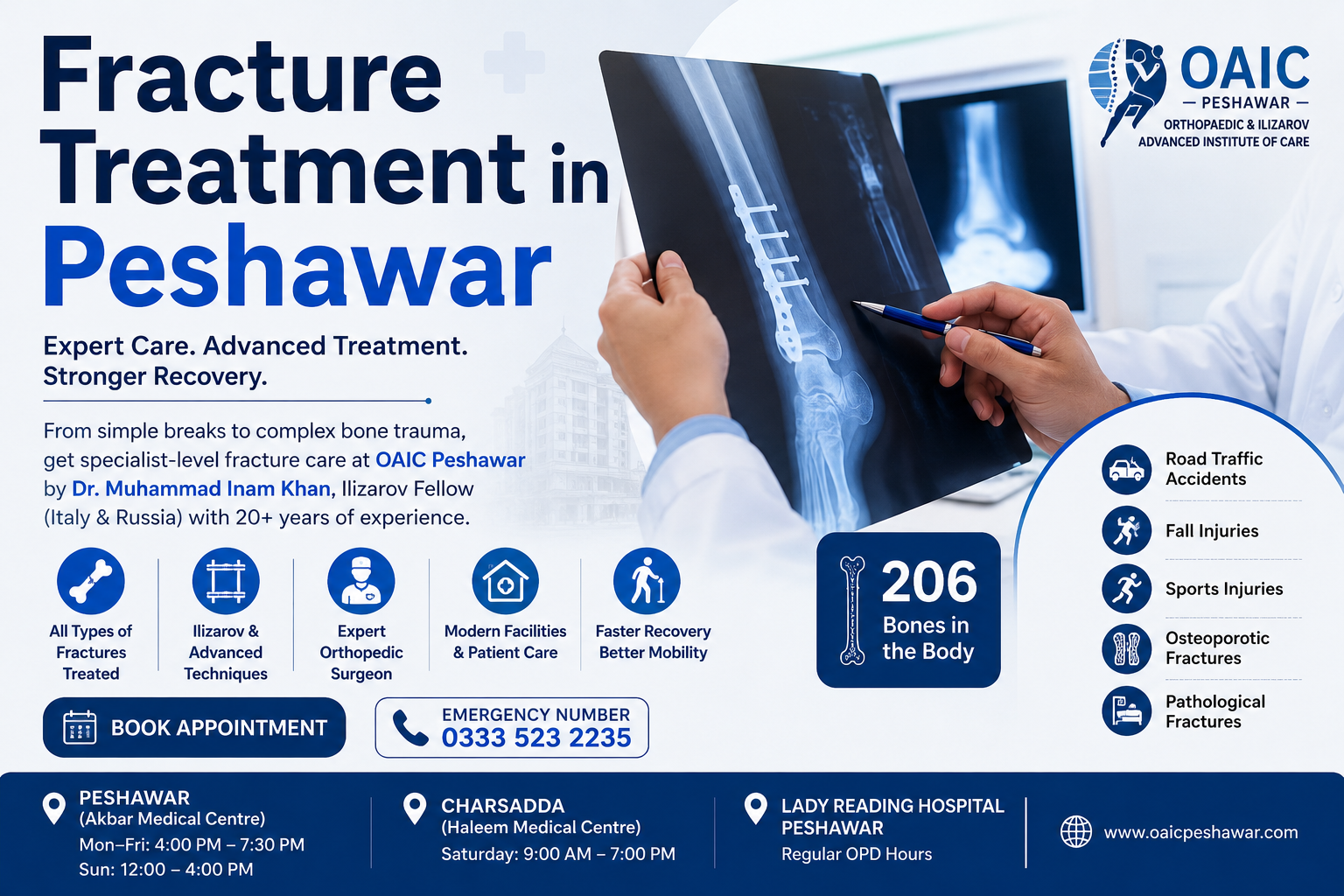
A broken bone demands immediate specialist care. At OAIC Peshawar, Dr Muhammad Inam Khan, fellowship-trained in the Ilizarov technique from Italy and Russia, provides expert Fracture Treatment in Peshawar, treating all fractures, from simple breaks to the most complex bone trauma. Whether caused by a road traffic accident, fall, sports injury, or weakened bones, you receive specialist-level fracture care at OAIC.
Common Fractures Treated at OAIC
Simple to Complex —
All Fractures Managed Here
A Bone Fracture, commonly known as a broken bone, occurs when physical force, trauma, or an underlying bone-weakening disease exceeds the strength of the bone. Fractures are one of the most common orthopaedic emergencies in Peshawar and across KPK, frequently caused by road traffic accidents, falls from height, sports injuries, and, in elderly patients, osteoporosis. Patients often experience severe fracture pain, swelling, and difficulty moving the affected limb.
There are over 20 distinct types of fractures, ranging from a simple, hairline crack (stress fracture) to a severely shattered bone with multiple fragments (comminuted fracture). Each type requires a different treatment approach, which is why specialist assessment by an experienced orthopaedic surgeon like Dr Muhammad Inam Khan is essential before any treatment begins.
No two fractures are the same. The bone affected, the fracture pattern, the degree of displacement, the patient’s age, activity level, and bone quality all influence the optimal treatment. At OAIC Peshawar, every fracture is evaluated individually from the imaging through to the final fixation method to ensure the fastest, safest, and most complete recovery possible through expert Fracture Treatment in Peshawar.
Dr Muhammad Inam Khan brings a uniquely high level of fracture expertise to Peshawar with advanced training in the Ilizarov technique (Italy and Russia) for complex fracture reconstruction, as well as standard fracture fixation techniques, including nailing, plating, and external fixation. He has treated thousands of fracture patients across KPK at OAIC and Lady Reading Hospital over 20+ years.
Peshawar (Akbar Medical Centre):
Mon–Fri: 4:00 PM – 7:30 PM · Sun: 12:00–4:00 PM
Charsadda (Haleem Medical Centre):
Saturday: 9:00 AM – 7:00 PM
Lady Reading Hospital, Peshawar:
Regular OPD Hours
Types of Fractures
Understanding your fracture type is the first step to the right treatment. Dr. Inam Khan diagnoses and manages all fracture patterns from the most routine to the most complex, ensuring accurate Bone Fracture Diagnosis.

The bone breaks but does not pierce the skin. The most common type is caused by falls, road accidents, or direct impact. It may be undisplaced (the bone remains aligned) or displaced (fragments have moved from their correct position), requiring accurate Bone Injury Evaluation. E.g., wrist fracture from fall, ankle fracture, clavicle fracture.
The bone shatters into three or more fragments. Usually caused by high-energy trauma such as road traffic accidents or serious falls. Almost always requires surgical fixation to reassemble the fragments and restore bone alignment through expert Orthopedic Trauma Care.
E.g., severe femur fracture, tibial plateau fracture in RTA.
A hairline crack in the bone caused by repeated stress rather than a single trauma. Common in athletes, soldiers, and young people who dramatically increase their activity level. Often misdiagnosed as a muscle strain or sprain, making early Bone Fracture Diagnosis essential.
E.g., metatarsal stress fracture, tibial stress fracture in runners.
The human body has 206 bones. Dr Inam Khan at OAIC Peshawar treats fractures across all regions from the most routine to the most surgically demanding.
The humerus, clavicle (collarbone), and shoulder blade are common in falls and sports, often requiring expert Orthopedic Assessment.
Very common in KPK
The humerus, clavicle (collarbone), and shoulder blade are common in falls and sports, often requiring prompt Bone Injury Evaluation.
Most common fracture
The humerus, clavicle (collarbone), and shoulder blade are common in falls and sports, often requiring prompt Bone Injury Evaluation.
Very common in KPK
The humerus, clavicle (collarbone), and shoulder blade are common in falls and sports, often requiring accurate Bone Fracture Diagnosis.
Very common in KPK
The humerus, clavicle (collarbone), and shoulder blade are common in falls and sports, often requiring comprehensive Orthopedic Examination.
Very common in KPK
The humerus, clavicle (collarbone), and shoulder blade are common in falls and sports, often requiring timely Bone Healing Management.
Very common in KPK
The humerus, clavicle (collarbone), and shoulder blade are common in falls and sports, often requiring specialized Bone Trauma Assessment.
Very common in KPK
The humerus, clavicle (collarbone), and shoulder blade are common in falls and sports, often requiring expert Musculoskeletal Injury Care.
Very common in KPK
Accurate imaging is essential; different fracture types require different investigations. Dr Inam Khan selects the right test for your specific fracture to plan the most effective treatment, ensuring accurate Bone Fracture Diagnosis.
01
Understanding exactly how the injury occurred—fall height, speed of impact, and direction of force—helps predict the fracture pattern and severity before any imaging is performed, enabling accurate Bone Trauma Assessment.
02
Assessment of the injury site for deformity, swelling, tenderness, neurovascular status (pulse, sensation, and movement), and skin integrity, including ruling out open fracture or compartment syndrome, is essential for a thorough Orthopedic Assessment.
03
The first-line investigation for all suspected fractures. Two views (front and side) of the injured bone are taken. It shows the fracture line, displacement, angulation, and bone quality. Fast and widely available, it plays a vital role in accurate Bone Fracture Diagnosis.
04
Computed tomography provides 3D imaging for complex or comminuted fractures, especially around joints (e.g., tibial plateau, calcaneus, pelvis), where precise surgical planning and accurate Bone Trauma Assessment are required before fixation.
05
Stress fractures are often invisible on X-rays initially. MRI detects early bone marrow stress reactions and confirms fractures before they become a complete break, making it essential for early Bone Injury Evaluation and return-to-sport decisions.
Mandatory for all fractures. It is fast, widely available and shows fracture line, displacement and angulation clearly. First-line investigation
3D imaging for complex, comminuted or intra-articular fractures. Essential for surgical planning of difficult injuries. Complex & joint fractures.
Detects occult (hidden) fractures, stress fractures, and bone marrow oedema not visible on X-ray. Stress & occult fractures
Pre-operative assessment, bone health markers (calcium, vitamin D, and bone density for osteoporotic fractures). Pre-surgery / elderly.
For pathological or osteoporotic fractures, measures bone mineral density to guide anti-osteoporosis treatment. Elderly people & osteoporosis.
Stress fractures are often invisible on X-rays initially. MRI detects early bone marrow stress reactions and confirms fractures before they become a complete break, making it essential for accurate Fracture Treatment in Peshawar and return-to-sport decisions.
Stress fractures are often invisible on X-rays initially. MRI detects early bone marrow stress reactions and confirms fractures before they become a complete break, making it essential for accurate Bone Trauma Assessment and return-to-sport decisions.
Rapid swelling around the injury site develops within minutes to hours of the fracture. Bruising (discolouration) often appears and spreads over 24–48 hours as blood from the fracture site tracks through the tissue, making early Bone Injury Evaluation essential.
The injured limb or bone may appear visibly bent, angled, rotated, or shortened compared to the normal side—a strong indicator of a displaced fracture requiring urgent Orthopedic Examination at OAIC.
Inability to move the affected limb normally, or to put any weight on a fractured leg or foot, is a significant clue, though some hairline fractures may allow partial movement initially and still require prompt Bone Injury Evaluation.
Inability to move the affected limb normally, or to put any weight on a fractured leg or foot, is a significant clue, though some hairline fractures may allow partial movement initially and still require a thorough Orthopedic Assessment.
If a bone is protruding through the skin or there is an open wound near the fracture site, this is a compound (open) fracture. This is a surgical emergency. Do not attempt to push the bone back. Seek immediate care for urgent Orthopedic Trauma Care.
Apply gentle pressure with a clean cloth if there is an open wound. Do not attempt to clean inside a wound.
If surgery may be needed, the patient should remain fasted.
Do not try to straighten the limb. Support it in the position found using padding, pillows, or a makeshift splint.
wrapped in cloth (not directly on skin) to reduce swelling. Do not apply heat.
If surgery may be needed, the patient should remain fasted.
Call OAIC Peshawar (+92 313 5735713) or proceed to the nearest hospital without delay.
Many undisplaced or minimally displaced fractures heal well without surgery. Our treatment includes plaster or fibreglass casts, slings, walker boots, and functional braces to keep the bone properly aligned while it heals naturally, supporting Bone Healing.
Plaster or fibreglass cast – Simple fractures of the wrist, ankle, foot, and clavicle.
Below-knee walker boot – Foot and ankle fractures.
Arm sling – Clavicle and undisplaced humeral fractures.
Functional brace – Certain tibial shaft fractures.
If a bone is protruding through the skin or there is an open wound near the fracture site, it is known as a compound (open) fracture. This is a surgical emergency that requires immediate medical attention. Do not attempt to push the exposed bone back into place, as this can increase the risk of infection and further damage. Seek immediate care from a qualified Orthopedic Surgeon for urgent trauma management and proper recovery.
The fractured bone is surgically exposed, repositioned (reduced) into correct anatomical alignment, and held firmly with plates and screws. Essential for fractures around joints, where precise restoration of the joint surface is critical to prevent arthritis. This procedure is performed by an experienced Orthopedic Surgeon to ensure proper alignment, stability, and recovery.
Locking plates and screws for metaphyseal and peri-articular fractures
Ankle fractures (malleolar fractures) — restoration of joint alignment
Wrist fractures (distal radius) — particularly in active patients
Tibial plateau fractures — joint surface reconstruction
Screws are placed into the bone above and below the fracture and connected to a rigid external frame outside the body. Used for open (compound) fractures, severely contaminated wounds, or as a temporary measure before definitive internal fixation once the soft tissue has recovered. This technique is an important part of Trauma Care for managing complex injuries and supporting safe recovery.
Open fractures with contaminated wounds — controls bone without implant in contaminated field
Temporary stabilisation in polytrauma patients (multiple injuries)
Periarticular fractures with severe swelling — “damage control orthopaedics”
Pelvic fractures with haemodynamic instability
OAIC's Specialist Expertise
Screws are placed into the bone above and below the fracture and connected to a rigid external frame outside the body. Used for open (compound) fractures, severely contaminated wounds, or as a temporary measure before definitive internal fixation once the soft tissue has recovered. This technique is an important part of Trauma Care for managing complex injuries and supporting safe recovery with advanced Fracture Treatment in Peshawar.
Open fractures with contaminated wounds — controls bone without implant in contaminated field
Temporary stabilisation in polytrauma patients (multiple injuries)
Periarticular fractures with severe swelling — “damage control orthopaedics”
Pelvic fractures with haemodynamic instability
Ilizarov Fellowship Italy & Russia
One of very few in KPK with this qualification
Fracture recovery is a journey, not just an event. Understanding what happens at each stage helps patients stay on track and avoid complications.
Days 1–7
Immediate Care
Weeks 2–6
Early Healing
Weeks 6–12
Active Rehab
Months 3–6
Return to Function
Months 6–12
Full Recovery
Leading pain specialist clinic in Peshawar. Expert treatment for all bone, joint and musculoskeletal pain conditions by Dr. Muhammad Inam Khan — certified pain management pecialist.

